
 Cardiovascular Physiology Concepts, 3rd edition textbook, Published by Wolters Kluwer (2021)
 Normal and Abnormal Blood Pressure, published by Richard E. Klabunde (2013)
Normal Heart Rhythm
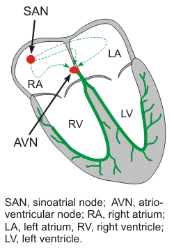 The rhythm of the heart is normally determined by a pacemaker site called the sinoatrial (SA) node located in the posterior wall of the right atrium near the superior vena cava. The SA node comprises specialized cells that undergo spontaneous generation of action potentials at 100-110 action potentials ("beats") per minute. This intrinsic rhythm is strongly influenced by autonomic nerves, with the vagus nerve being dominant over sympathetic influences at rest. This "vagal tone" brings the resting heart rate down to 60-70 beats/minute. The normal range for sinus rhythm is 60-100 beats/minute. Sinus rates below this range are termed sinus bradycardia, and sinus rates above this range are termed sinus tachycardia.
The sinus rhythm normally controls both atrial and ventricular rhythm. Action potentials generated by the SA node spread throughout the atria, depolarizing this tissue and causing atrial contraction. The impulse then travels into the ventricles via the atrioventricular node (AV node). Specialized conduction pathways (bundle branches and Purkinje fibers) within the ventricle rapidly conduct the wave of depolarization throughout the ventricles to elicit ventricular contraction. Therefore, normal cardiac rhythm is controlled by the pacemaker activity of the SA node.
Abnormal cardiac rhythms can occur if:
- the SA node does not function normally (e.g., sinus bradycardia or tachycardia).
- impulses are not conducted from the atria to the ventricles through the AV node (termed AV block).
- abnormal conduction pathways are followed (e.g., accessory pathways between atria and ventricles).
- other pacemaker sites within the atria or ventricles (e.g., ectopic pacemakers) trigger depolarization.
Revised 10/27/2023