Cardiac Cycle - Isovolumetric Relaxation (Phase 5)
All Valves Closed

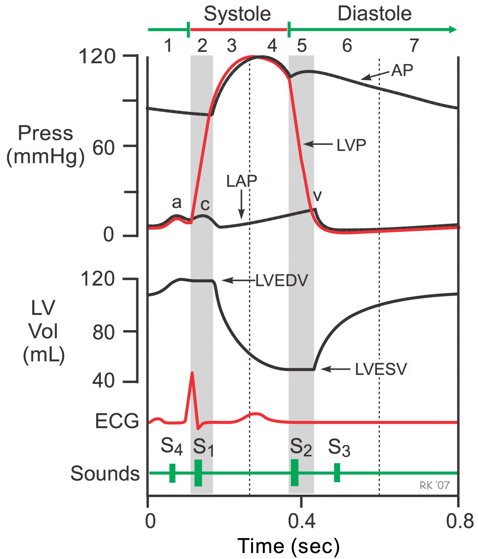
 When the intraventricular pressures fall sufficiently at the end of phase 4, the aortic and pulmonic valves abruptly close (aortic precedes pulmonic) causing the second heart sound (S2) and the beginning of isovolumetric relaxation. Valve closure is associated with a small backflow of blood into the ventricles and a characteristic notch (incisura or dicrotic notch) in the aortic and pulmonary artery pressure tracings.
After valve closure, the aortic and pulmonary artery pressures rise slightly (dicrotic wave) followed by a slow decline in pressure.
The rate of pressure decline in the ventricles is determined by the rate of relaxation of the muscle fibers, which is termed lusitropy. This relaxation is regulated largely by the sarcoplasmic reticulum, that are responsible for rapidly re-sequestering calcium following contraction (see excitation-contraction coupling).
Although ventricular pressures decrease during this phase, volumes do not change because all valves are closed. The volume of blood that remains in a ventricle is called the end-systolic volume and is ~50 mL in the left ventricle. The difference between the end-diastolic volume and the end-systolic volume is ~70 ml and represents the stroke volume.
Left atrial pressure (LAP) continues to rise because of venous return from the lungs. The peak LAP at the end of this phase is termed the v-wave.
Jump to other phases:
- Phase 1 - Atrial Contraction
- Phase 2 - Isovolumetric Contraction
- Phase 3 - Rapid Ejection
- Phase 4 - Reduced Ejection
- Phase 6 - Rapid Filling
- Phase 7 - Reduced Filling
Revised 11/04/2023

 Cardiovascular Physiology Concepts, 3rd edition textbook, Published by Wolters Kluwer (2021)
 Normal and Abnormal Blood Pressure, published by Richard E. Klabunde (2013)