
 Cardiovascular Physiology Concepts, 3rd edition textbook, Published by Wolters Kluwer (2021)
 Normal and Abnormal Blood Pressure, published by Richard E. Klabunde (2013)
Ion Channels
The cell membrane is permeable to several ions, the most important of which are Na+, K+, Ca++, and Cl-. These ions pass across the membrane through specific ion channels that can open (become activated) and close (become inactivated). Therefore, these channels are said to be gated channels. Their opening and closing can occur in response to
- voltage changes (voltage gated channels)
- ligand activation of receptors (receptor gated channels)
- specific ions and chemical ligands
Cardiac cells can have multiple channels for a particular ion. For example, there are many types of potassium channels that play an essential role in resting membrane potential and in action potentials. It is the opening and closing of ion channels that alters specific ion conductances in a manner that determines resting potentials and generates action potentials. For example, when an action potential is elicited in a cardiomyocyte, sodium channels transiently open and potassium channels close, which leads to depolarization. Shortly thereafter (within a few milliseconds), the sodium channels close and calcium channels open to maintain a depolarized state. This is followed by inactivation of the calcium channels and a reopening of the potassium channels, which leads to membrane repolarization.
The following table summarizes some important ion channels that are found in cardiac and vascular smooth muscle cells:
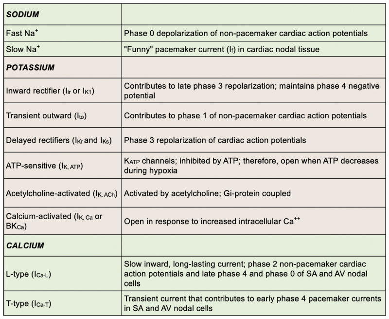
Many of the antiarrhythmic drugs that are used to treat cardiac arrhythmias have their action on sodium, calcium, and potassium channels.
Revised 11/02/2023