
 Cardiovascular Physiology Concepts, 3rd edition textbook, Published by Wolters Kluwer (2021)
 Normal and Abnormal Blood Pressure, published by Richard E. Klabunde (2013)
Cardiac Anatomy
The detailed anatomy of the heart can be found in anatomy textbooks. The following presents only a brief description of cardiac anatomy so that the physiology of the cardiac cycle can be understood.
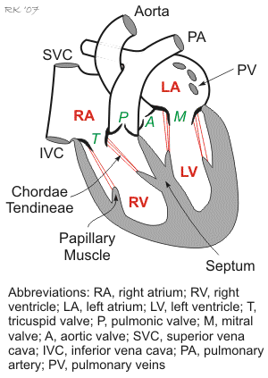 Venous blood enters the right atrium (RA) of the heart through the superior vena cava (SVC) and inferior vena cava (IVC). The right atrium has a relatively thin muscular wall and easily expands with blood as it fills (i.e., it is highly compliant). Because of its high compliance, the RA pressure is normally very low (0-3 mmHg). It also undergoes spontaneous contractions (see cardiac cycle) to aid in the filling of the right ventricle (RV). Blood passes from the RA to the RV through the tricuspid valve. The free wall of the right ventricle is not as thick as the left ventricle, and anatomically, it wraps itself around part of the larger, and thicker, left ventricle. The RV wall, however, is thicker and more muscular than the RA, so that when it contracts, it can develop considerably more pressure (~25 mmHg) than the RA. As the RV contracts and generates pressure, blood leaves the RV, flows across an open semilunar pulmonic valve, and enters the pulmonary artery that distributes the output of the right ventricle to the lungs where exchange of oxygen and carbon dioxide occurs. The pulmonic valve, like all healthy heart valves, permits blood to flow in only one direction. Blood returns to the heart from the lungs through four pulmonary veins that enter the left atrium (LA). This chamber, like the RA, has thin walls and is very distensible; however, the blood pressure within the LA is several mmHg higher than the RA pressure (6-10 mmHg in the LA compared to 0-3 mmHg in the RA). Blood flows from the LA, across the mitral valve, and into the left ventricle (LV). The LV wall is very thick so that it can generate high pressures when it contracts (normally ~120 mmHg at rest). When the LV contracts, it expels blood through the semilunar aortic valve and into the aorta, which then distributes blood to the arterial system.
Tricuspid and mitral valves (also called atrioventricular, or AV valves) have fibrous strands (chordae tendineae) on their leaflets that attach to papillary muscles located on the respective ventricular walls. The papillary muscles contract during ventricular contraction and generate tension on the valve leaflets via the chordae tendineae to prevent the AV valves from bulging back into the atria and becoming incompetent. The semilunar valves (pulmonic and aortic) do not have analogous attachments.
Continue to Cardiac Cycle
Mini-Lecture: Cardiac Functional Anatomy (3 minutes) (CLICK HERE)
Mini-Lecture: Normal Intracardiac Pressures (3 minutes) (CLICK HERE)
Revised 11/04/2023