Cardiac Valve Disease
- What are heart valves, and what is their function?
- What causes valve dysfunction?
- How are valve defects classified?
- What are the consequences and clinical symptoms of valve disease?
What are heart valves, and what is their function?
Valves within the heart separate the right atrium and ventricle (tricuspid valve), the left atrium and ventricle (mitral valve), the right ventricle and the pulmonary artery (pulmonic valve), and the left ventricle and aorta (aortic valve) (click here to see cardiac anatomy diagram). The valves ensure that blood flows in a single pathway through the heart by opening and closing in a particular time sequence during the cardiac cycle. Normal valves permit blood to flow in only one direction, for example, from the right atrium into the right ventricle. When heart valves become diseased or damaged, they may not fully open or close. This can seriously impair cardiac function by causing blood to leak backwards into cardiac chambers or by requiring heart chambers to contract more forcefully to move blood across a narrowed valve.
What causes valve dysfunction?
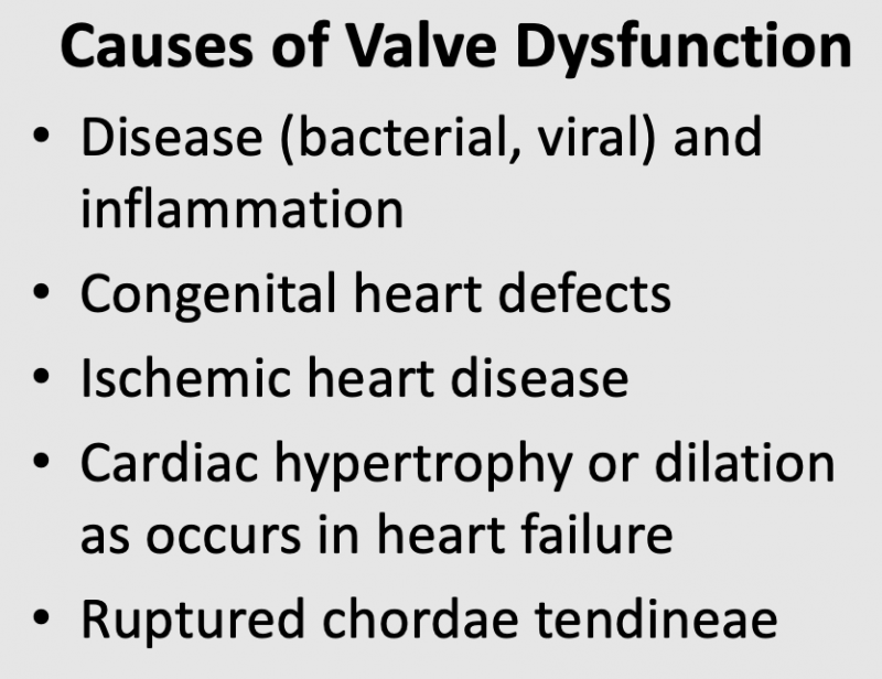
 A chronic disease process is responsible for defective valves in older individuals. Sometimes, the disease results from an event many years earlier, such as rheumatic fever. Bacterial infection, viral infection and inflammation of valves (infective endocarditis) can produce changes in valve structure and function. Normally, valve leaflets are very thin and flexible, but they can become thickened and rigid in response to disease processes. When this occurs, the valve leaflets may not fully open or completely close. Valve disease found in younger individuals is usually because of a congenital defect in the embryological development of the heart.
Valve dysfunction can occur secondarily to other cardiac diseases, such as coronary artery disease, cardiac hypertrophy, and cardiac dilation. If coronary artery disease progresses to where papillary muscles become hypoxic or infarcted, then the impaired contractile function of these muscles can lead to a leaky tricuspid or mitral valve. Cardiac hypertrophy or dilation, by altering cardiac chamber structure and dimensions, can lead to valve dysfunction. Finally, valve dysfunction can also occur if the chordae tendineae connecting the valve leaflet to the papillary muscle rupture.
How are valve defects classified?
There are two general types of cardiac valve defects: stenosis and insufficiency. Some patients, however, may have a combination of stenosis and insufficiency. Valvular stenosis results from a narrowing of the valve orifice (reduced cross-sectional area of the opened valve) that is usually caused by a thickening and increased rigidity of the valve leaflets, often accompanied by calcification. When this occurs, the valve does not open completely as blood flows across it, resulting in a high resistance to flow and the development of a large pressure gradient across the valve when blood is flowing through the valve. Pressure increases in the chamber proximal to the valve and decreases in the chamber or artery distal to the valve.
Valvular insufficiency occurs when the valve leaflets do not completely seal when the valve is closed. This causes regurgitation of blood (backward flow of blood) into the proximal chamber. For example, in aortic valve insufficiency, blood regurgitates from the aorta back into the left ventricle after ventricular ejection.
What are the clinical symptoms of defective valves?
Valvular stenosis and insufficiency can have serious cardiac consequences, and produce the following clinical symptoms:
- Shortness of breath (dyspnea)
- Fatigue
- Reduced exercise capacity
- Light-headedness or fainting (syncope)
- Heart failure
- Pulmonary hypertension
- Pulmonary/systemic edema
- Chest pain (angina)
- Arrhythmias
- Blood clots (thromboembolism) which can cause stroke
Revised 01/26/2023

 Cardiovascular Physiology Concepts, 3rd edition textbook, Published by Wolters Kluwer (2021)
 Normal and Abnormal Blood Pressure, published by Richard E. Klabunde (2013)