Microcirculation Structure and Function
The microcirculation comprises arterioles, capillaries, venules, and terminal lymphatic vessels.
Arterioles
- Small precapillary resistance vessels (10-200 μ) composed of an endothelium surrounded by one or more layers of smooth muscle cells
- Richly innervated by sympathetic adrenergic fibers and highly responsive to sympathetic vasoconstriction via both α1 and α2 postjunctional receptors.
- Represent the major site for regulating systemic vascular resistance.
- Rhythmical contraction and relaxation of arterioles sometimes occurs (i.e., spontaneous vasomotion).
- Primary function of arterioles within an organ is flow regulation, which determines oxygen delivery and the washout of metabolic by-products from the tissue.
- Regulate, in part, capillary hydrostatic pressure and therefore influence capillary fluid exchange.
Capillaries
- Small exchange vessels (6-10 μ) composed of highly attenuated (very thin) endothelial cells surrounded by basement membrane − no smooth muscle.
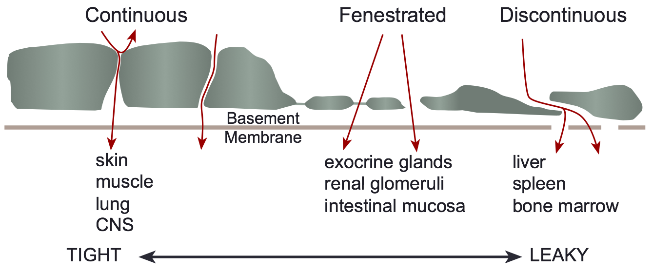
 Three structural classifications:
- Continuous (found in muscle, skin, lung, central nervous system) − basement membrane is continuous and intercellular clefts (between adjacent endothelial cells) are tight (i.e., have tight junctions); these capillaries have the lowest permeability.
- Fenestrated (found in exocrine glands, renal glomeruli, intestinal mucosa) − perforations (fenestrae) in endothelium result in relatively high permeability.
- Discontinuous (found in liver, spleen, bone marrow) − large intercellular gaps and gaps in basement membrane result in extremely high permeability.
- Large surface area and relatively high permeability (especially at intercellular clefts) to fluid and macromolecules make capillaries the primary site of exchange for fluid, electrolytes, gases, and macromolecules.
- In some organs, the number of perfused capillaries can increase during high flow states (e.g., in contracting muscle) in response to arteriolar vasodilation and redistribution of precapillary pressures
Venules
- Small exchange vessels (10-200 μ) composed of endothelial cells surrounded by basement membrane (smallest postcapillary venules) and smooth muscle (larger venules).
- Fluid and macromolecular exchange occur at small postcapillary venular junctions.
- Sympathetic innervation of larger venules can alter venular tone, which plays a role in regulating capillary hydrostatic pressure.
Terminal Lymphatics
- Composed of endothelium with intercellular gaps surrounded by highly permeable basement membrane and are similar in size to venules − terminal lymphatics end as blind sacs.
- Larger lymphatics also have smooth muscle cells.
- Spontaneous and stretch-activated vasomotion is present, which serves to pump lymph out of the tissue.
- Sympathetic nerves can modulate vasomotion and cause contraction.
- One-way valves direct lymph away from the tissue and eventually back into the systemic circulation via the thoracic duct and subclavian veins (2-4 liters/day returned).
Revised 11/06/2023

 Cardiovascular Physiology Concepts, 3rd edition textbook, Published by Wolters Kluwer (2021)
 Normal and Abnormal Blood Pressure, published by Richard E. Klabunde (2013)