
 Cardiovascular Physiology Concepts, 3rd edition textbook, Published by Wolters Kluwer (2021)
 Normal and Abnormal Blood Pressure, published by Richard E. Klabunde (2013)
Cardiac Signal Transduction Mechanisms (G-Protein-Linked)
There are several major signal transduction mechanisms found in cells of the cardiovascular system, the most important being the G-protein and nitric oxide-cyclic GMP pathways. Described below are the G-protein-coupled pathways found in the heart. Signal transduction mechanisms regulating vascular smooth muscle contraction and relaxation are found elsewhere (Click Here).
Gs-Protein and Gi-Protein Coupled Signal Transduction
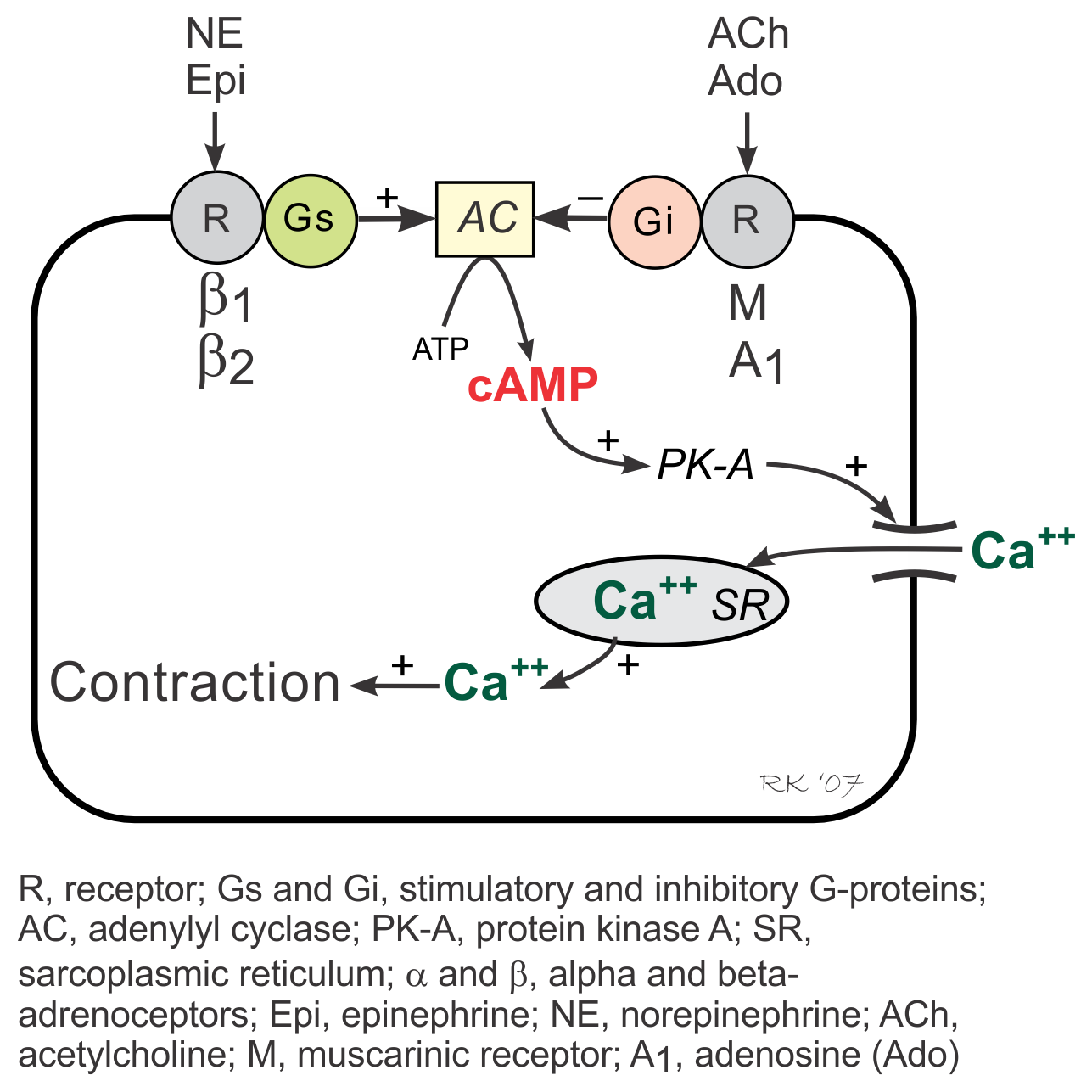 G-proteins are linked to an enzyme, adenylyl cyclase, that dephosphorylates ATP to form cyclic AMP (cAMP). Gs-protein (stimulatory G-protein) activation (e.g., via β-adrenoceptors) increases cAMP by activating adenylyl cyclase. cAMP then activates PK-A (cAMP stimulated protein kinase) and causes increased cellular influx of Ca++ by phosphorylating and activating membrane L-type calcium channels, and by enhancing release of Ca++ by the sarcoplasmic reticulum in the heart. These and other intracellular events increase inotropy (muscle contractility), chronotropy (heart rate), dromotropy (velocity of electrical conduction) and lusitropy (relaxation rate).
Activation of Gi-proteins (inhibitory G-protein), for example by adenosine and muscarinic agonists (e.g., acetylcholine) binding to their receptors, decreases cAMP (through adenylyl cyclase inhibition), inactivates PK-A, decreases Ca++ entry into the cell and release by the sarcoplasmic reticulum, and increases outward, hyperpolarizing K+ currents.
Gi-protein activation produces effects that are opposite to those elicited by Gs-protein activation. Because Gi-protein effects are primarily found in the SA node and AV node where there are an abundance of Gi-protein coupled receptors, activation of this pathway leads to a decrease in sinus rate and AV nodal conduction velocity with minimal effects on muscle contractility, particularly in the ventricles. In contrast, Gs-protein strongly stimulates muscle contraction, besides having nodal effects.
 Gq-Protein and IP3- Coupled Signal Transduction
The IP3 pathway is linked to activation of α1-adrenoceptors, angiotensin II (AII) receptors, and endothelin-1 (ET-1) receptors and therefore is stimulated by alpha-agonists, angiotensin II and endothelin-1. These receptors are coupled to a phospholipase C (PL-C)-coupled Gq-protein, which when activated, stimulates the formation of inositol triphosphate (IP3) from phosphatidylinositol biphosphate (PIP2). Increased IP3 stimulates Ca++ release by the sarcoplasmic reticulum in the heart, increasing inotropy as one of its actions. Gq-protein coupled responses in the heart, however, are relatively minor compared to Gs-protein coupled responses.
Altered Signal Transduction in Heart Disease
Altered signal transduction mechanisms have a significant role in the loss of inotropy in heart failure. For example, desensitization of β1-adrenoceptors in the heart decreases inotropic responses to sympathetic activation. Uncoupling of the β1-adrenoceptor and the Gs-protein reduces the ability to activate adenylyl cyclase. If the ability of protein kinase A to phosphorylate L-type calcium channels is impaired, then calcium influx into the cell is reduced, leading to a smaller release of calcium by the sarcoplasmic reticulum. Reduced calcium release impairs excitation-contraction coupling, decreasing inotropy.
Revised 12/7/2022